Back

Select your region

Schedule a cord blood consultation

Make an appointment
Problems of a newborn born prematurely

Problems of a newborn born prematurely

14.01.2023

5 mins of reading

Kinga Żebrowska

Kinga Żebrowska

Graduate of Warsaw Medical University

Premature babies, as babies born before 37 are referred to. week of pregnancy, they often have to face many difficulties from the first days of their lives. Low birth weight and possible complications, cause many Parents to fear for their baby's life and health. What are the most common complications of prematurity and how does modern medicine deal with them?

Newborns born prematurely are a very broad group including:

  • extreme prematurity, or those born before 28. week of pregnancy,
  • late ones who were born between 34. a 36. week.

The number and severity of complications associated with prematurity depend mainly on the baby’s birth weight and gestational age. The capabilities and medical facilities of the hospital where the birth takes place are also not insignificant. The most common complications in premature babies are respiratory distress, bronchopulmonary dysplasia (BPD) and neurological problems. In the text, we focus on three selected problems of newborns born prematurely – persistent ductus arteriosus, necrotizing enterocolitis and retinopathy of prematurity.

Persistent ductus arteriosus

A patent ductus arteriosus (PDA) occurs in 20-30% of premature newborns.

The Botall’s duct is a vessel that connects the pulmonary trunk to the aorta during fetal life. It is made up of smooth muscle cells that are particularly sensitive to prostaglandins and oxygen. During fetal life, prostaglandins secreted by the placenta keep the ductus arteriosus open, and after birth, when the concentration of oxygen in the blood increases, it shrinks.

The arterial duct usually closes in the first few days of a newborn’s life. If this does not happen, there is a leakage of blood from the systemic circulation to the pulmonary circulation. This carries the consequences of overloading the heart, as well as impeding gas exchange in the alveoli of the newborn’s lungs.

Sometimes it is difficult to diagnose a persistent ductus arteriosus due to the lack of clear symptoms. One of them is the systolic-diastolic murmur during auscultation of the baby’s heart, which the doctor can detect during a routine examination. Infants may also experience episodes of cyanosis, apnea or bradycardia (a conditionwhen the number of heartbeats falls below 60 per minute).

When a Botall’s duct is diagnosed in a premature infant, drug treatment is attempted, and the drugs administered are aimed at closing it. If this action does not have the desired effect, a PDA shutdown operation is performed. Lack of appropriate treatment leads to the development of pulmonary hypertension, as well as right-left leakage and Eisenmenger syndrome. However, there are exceptions when a persistent ductus arteriosus should not be closed until the other defect is cured. This happens in the case of, among others. conduction-related heart defects.

Srodtext Course v1 1

Necrotizing enterocolitis

Necrotizing enterocolitis (NEC- necrotizing enterocolitis) affects about 7% of newborns born before 32. week of pregnancy. Its etiology is not fully understood, but the most important risk factors are hypoxia and bacterial invasion.

Necrotizing enterocolitis usually appears around 2. A week in the life of a newborn. It manifests itself in various ways and is most often characterized by a turbulent course. The child’s condition rapidly deteriorates, sometimes accompanied by symptoms of septic shock. In many cases, Parents also notice food intolerance and blood in the child’s stool. His abdomen, on the other hand, is hard and tender.

To diagnose necrotizing enterocolitis, an X-ray or ultrasound is usually performed, which shows bowel dilatation. The lack of peristalsis is also characteristic.

The cornerstone of treatment is the abandonment of enteral (intragastric and enteral) feeding. Intravenous antibiotics, fluids are administered and an adequate supply of calories is provided. If these measures are unsuccessful or there is a perforation of the intestine, surgical intervention is required. A distant complication of necrotizing enterocolitis surgery can be short bowel syndrome.

Retinopathy of prematurity

Retinopathy of prematurity  (ROP – retinopathy of prematurity) is an abnormal development of retinal vascularization, which is directly related to the immaturity of the child and the tissues that build his eye.

Causes of retinopathy include oxygen treatment (oxygen therapy), hyperglycemia, anemia and coagulation disorders in the newborn. Maternal diseases during pregnancy such as hypertension, preeclampsia (pre-eclampsia) or diabetes are also risk factors.

It is very important to perform systematic ophthalmological follow-up in premature infants until about 3-4 years of age until the retina reaches maturity. Vascular lesions can regress spontaneously or, on the contrary, untreated, even lead to retinal detachment and blindness. The most common treatment is laser therapy, which has an estimated effectiveness of approx. 90%.

Can cord blood be collected in premature babies?

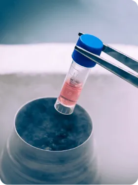
The procedure for collecting cord blood looks the same for a natural childbirth as for a cesarean section. Gestational age is also irrelevant here, so prematurity does not preclude stem cell storage. What’s more – complications of prematurity can be treated with banked material. Stem cells are used, among other things, in patients with cerebral palsy, of which prematurity is a risk factor.

Stem cells from cord blood are among the multipotent cells. This means that they have the ability to transform into specialized cells such as bone, cartilage or fat cells. As a result, the list of conditions for which the potential of cord blood-derived cells is exploited is growing longer every year.

Cord blood is collected from the vessels of the umbilical cord and placenta after delivery. If the parents choose not to collect and store the stem cells, it is irretrievably disposed of, and with it disappears the chance to use the cells’ capabilities in the future in case of a possible disease in the child. More details: https://www.pbkm.pl/komorki-macierzyste/komorki-macierzyste-i-krew-pepowinowa

Rate this article:

Średnia ocen 0 / 5. Liczba głosów: 0

(
0
(0)
)

You may also be interested in