-
-
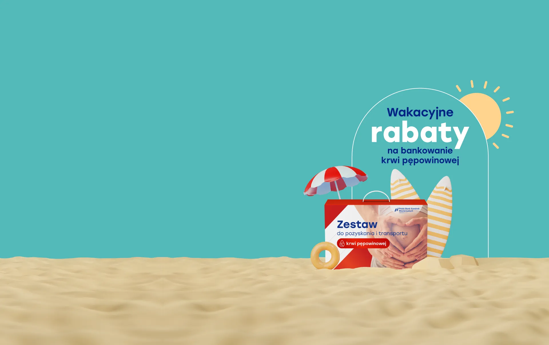
Donate cord blood for 1zł
and get discounts on other fees!
-
Win free banking for 18 years
or a voucher worth 2,000 PLN
Winners are selected every week!
-
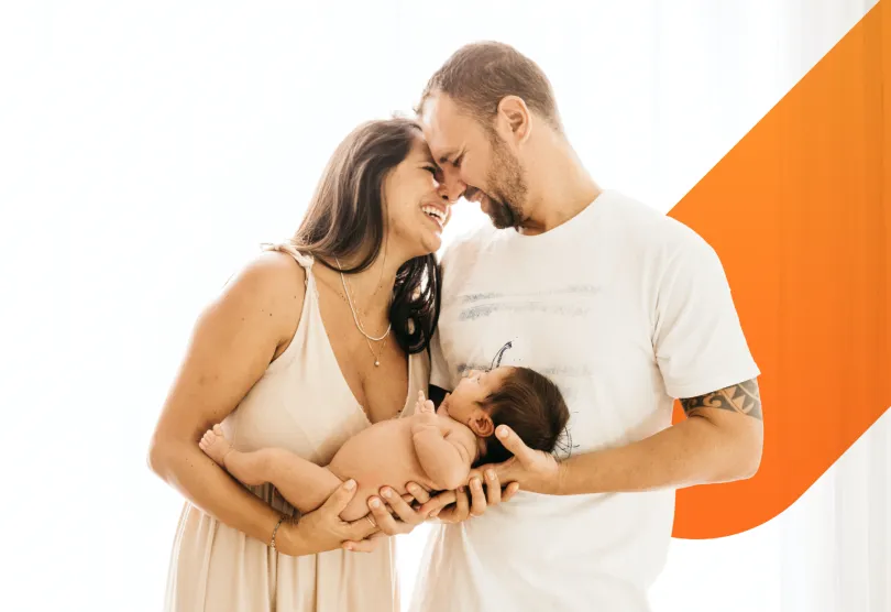
Secure your child’s future.
Invest in cord blood banking!
Get an extra chance to treat as many as 80 serious diseases.
-
Webinars for parents
Gain exceptional knowledge.
Already more than 85,000.
people have prepared for childbirth with our webinars.
Join them!
-
Prepare for childbirth
Free access to the video course Gain knowledge with our experts in the comfort of your home.
Already nearly 15,000.
people have benefited from our course.


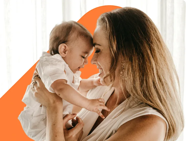
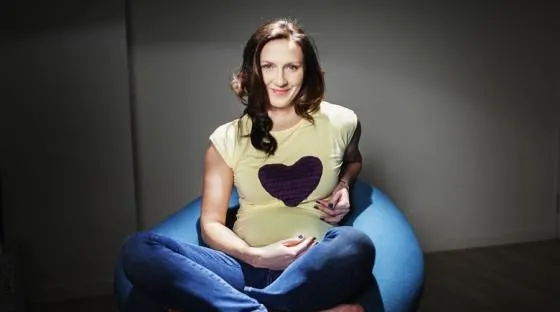
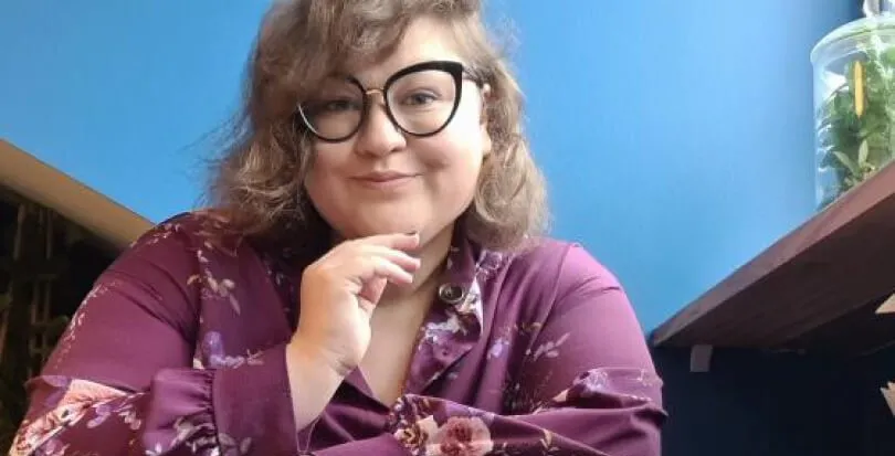
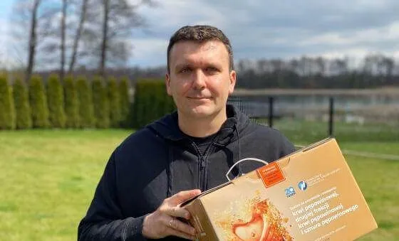
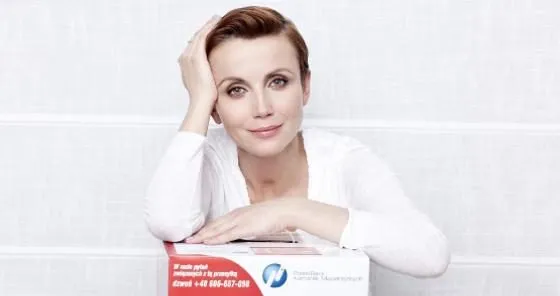
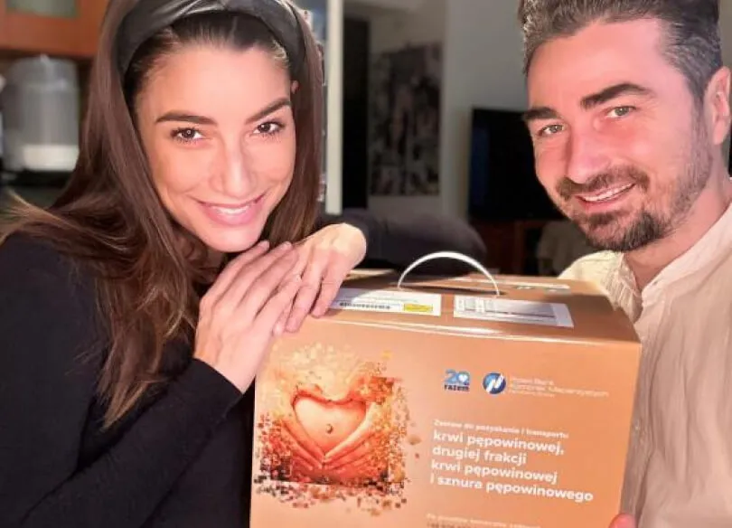
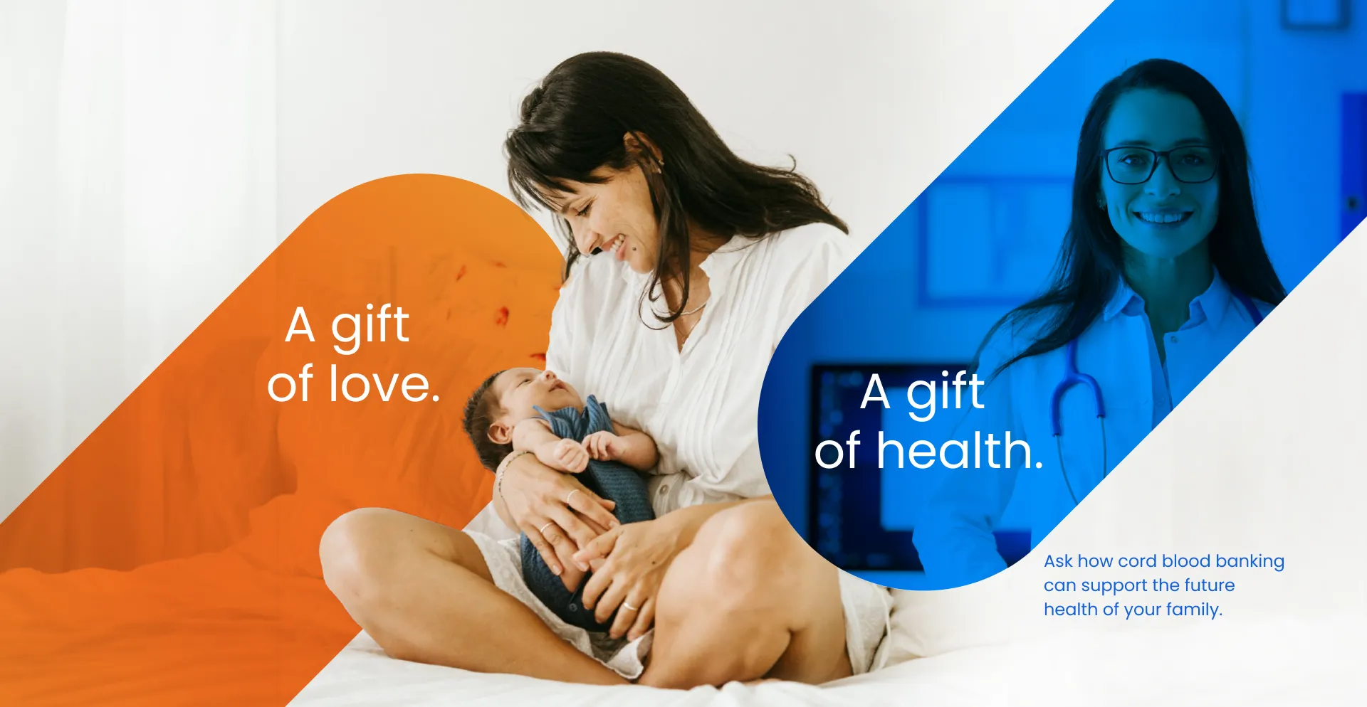
The gift of love
Securing umbilical cord blood at birth can provide your child with access to potential stem cell-based therapies in the future.
To learn more about the opportunity offered by stem cells, explore the stories of Parents who have secured cord blood.

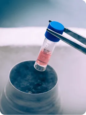
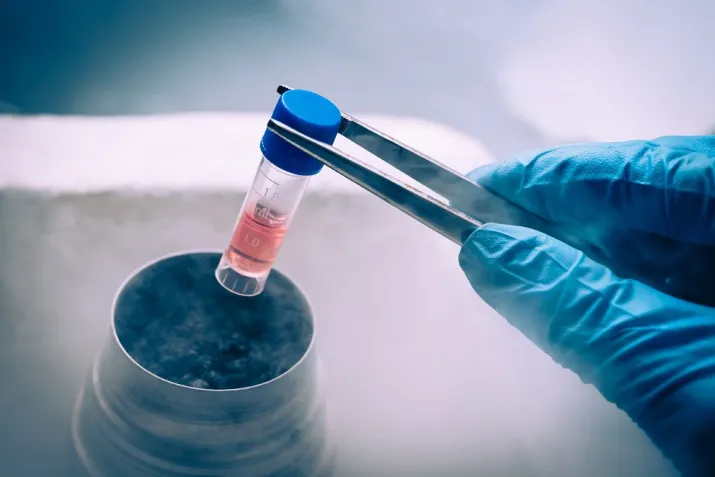
The gift of science
Cord blood stem cells, analogous to bone marrow stem cells, are used in the treatment of nearly 80. Severe oncological and hematological diseases. Research is underway on its use in autoimmune and neurological diseases.
Check out where they have applications.
PBKM in numbers
1 000 000
stored samples
1 500
cooperating hospitals
7 130
Patients have benefited from stem cell therapy
235
use your own cord blood